Advanced Analytical Centre Analytical Facilities All Instruments Laser Scanning Confocal Microscopy (LSCM)
Laser Scanning Confocal Microscopy (LSCM)
- MRCMHR
- Future Students
- JCU Global Experience
- International Students
- Student experience
- Open Day
- How to apply
- Pathways to university
- Living on Campus
- Courses
- Publications
- Mature students
- Scholarships
- Entry options
- JCU Families
- JCU Heroes Programs
- Aboriginal and Torres Strait Islander in Marine Science
- Elite Athletes
- Defence
- Capability.Co
- AI@JCU
- AALL
- Current Students
- Student Ambassador Program
- New students
- JCU Orientation
- LearnJCU
- Placements
- EDQS
- Unicare Centre and Unicampus Kids
- Graduation
- Off-Campus Students
- JCU Job Ready
- Safety and Wellbeing
- JCU Prizes
- Professional Experience Placement
- Employability Edge
- Art of Academic Writing
- Art of Academic Editing
- Careers and Employability
- Health, Wellbeing and Belonging
- Career Ready Plan
- Careers at JCU
- Partners and Community
- School Outreach and Widening Participation
- Alumni
- International partnerships
- About JCU
- Reputation and Experience
- Chancellery
- Governance
- Celebrating 50 Years
- Academy
- Indigenous Engagement
- Education Division
- Research Festival
- Graduate Research School
- Research Division
- Research and Innovation Services
- CASE
- College of Business, Law and Governance
- College of Healthcare Sciences
- College of Medicine and Dentistry
- College of Science and Engineering
- MPE
- Anthropological Laboratory for Tropical Audiovisual Research (ALTAR)
- Rural Remote and Tropical Health Systems
- Agriculture Technology and Adoption Centre (AgTAC)
-
Advanced Analytical Centre
- About us
- Commercial and external clients
-
Analytical Facilities
-
All Instruments
- X-ray Powder Diffraction (XRD)
- X-ray Fluorescence (XRF)
- Scanning Electron Microscopy (SEM)
- Inductively Coupled Plasma-Mass Spectrometer (ICP-MS)
- Laser ablation
- Inductively Coupled Plasma-Atomic Emission Spectrometer (ICP-AES)
- Gas Chromatography-Liquid Chromatography (GC/LC)
- Additional Equipment
- Laser Scanning Confocal Microscopy (LSCM)
- Advanced Analytical Centre
- Multicollector-Inductively Coupled Plasma-Mass Spectrometer (MC-ICP-MS)
- Electron Probe Microanalyser (EPMA or Microprobe)
- Sample Requirements
- Techniques and Facilities
-
All Instruments
- Staff
- Safety
-
Resources
- Element-to-stoichiometric oxide conversion factors
- Gunshot residue (GSR)
- FAQ ICP
- FAQ Organic
- SEM images of local insects – N.Queensland
- False coloured images
- Notes on sample preparation of biological material for SEM
- Routine XRF element analysis @ AAC
- How to view/edit element maps from Jeol 8200 EPMA
- Standard ICP element analysis @ AAC
- FAQ - XRD/XRF
- Other JCU Facilities
- Contact the AAC
- AMHHEC
- Aquaculture Solutions
- AMHRA
- JCU Digital Wellbeing Group
- ARCSTA
- Lions Marine Research Trust
- Australian Tropical Herbarium
- Australian Quantum & Classical Transport Physics Group
- Boating and Diving
- Clinical Psychedelic Research Lab
- Centre for Tropical Biosecurity
- Centre for Tropical Bioinformatics and Molecular Biology
- CITBA
- CMT
- Centre for Disaster Solutions
- CSTFA
- Cyclone Testing Station
- The Centre for Disaster Studies
- Daintree Rainforest Observatory
- Fletcherview
- JCU Eduquarium
- JCU Turtle Health Research
- MARFU
- Orpheus
- TESS
- JCU Ideas Lab
- CADSI
- CNL
- TARL
- eResearch
- Indigenous Education and Research Centre
- Past Course and Subject Handbooks
- Estate
- Work Health and Safety
- Staff
- Discover Nature at JCU
- Cyber Security Hub
- Association of Australian University Secretaries
- Services Division
- Environmental Research Complex [ERC]
- Foundation for Australian Literary Studies
- Gender Equity at JCU
- Give to JCU
- Indigenous Legal Needs Project
- Inherent Requirements
- IsoTropics Lab
- IT Services
- JCU Webinars
- JCU Events
- JCU Motorsports
- JCU Sport
- Library
- Mabo Decision: 30 years on
- Marine Geophysics Laboratory
- Office of the Vice Chancellor and President
- Outstanding Alumni
- Policy
- PAHL
- Queensland Research Centre for Peripheral Vascular Disease
- Rapid Assessment Unit
- RDIM
- Researcher Development Portal
- Roderick Centre for Australian Literature and Creative Writing
- Contextual Science for Tropical Coastal Ecosystems
- State of the Tropics
- Strategic Procurement
- Student profiles
- SWIRLnet
- TREAD
- TropEco for Staff and Students
- TropCOVID: Mapping COVID-19 Immunity in Far North Queensland
- TUDLab
- VAVS Home
- WHOCC for Vector-borne & NTDs
- Media
- Copyright and Terms of Use
- Australian Institute of Tropical Health & Medicine
- JCU Respect
- Pay review
- Research
If you have used this instrument please cite using the following:
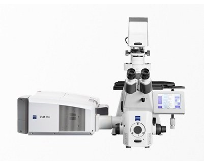
Zeiss. (2025). Laser Scanning Confocal Microscope (Zeiss LSM710). James Cook University, Advanced Analytical Centre. https://doi.org/10.25903/YTKP-AB62
Technique in brief
The confocal is a microscope which focusses only on a single focal plane. Lasers are used for point illumination and the resulting fluorescence is detected by a photodetector. The design of a confocal ensures that only light produced by fluorescence close to the focal plane is observed. By scanning across the sample line by line an image may be produced.
One of the advantages of a confocal microscope over a normal one is the ability to shift focal planes. Successive images generated at different focal plans can be combined to produce 3D images.
Current instrumentation
The current LSCM at the AAC is a Zeiss 710 with and inverted sytle microscope stage and 5 lasers available at the following wavelengths:
405nm
488nm
514nm
561nm
633nm
Applications
Confocal microscopy is used mainly but not exclusively for biological and life science applications. In general a variety of specific fluorescent stains or fluorphores are applied to a thin sections of biological material however other opaque and reflective materials can be observed given the right conditions.
Sample requirements
In general sample must be prepared for an inverted microscope, for biological samples a sealed cover slip must be used.
Considerations should include: Coverslip thickness and spacers, the opaque nature of the sample, sample thickness, mounting media and fluorescent probe application.