Advanced Analytical Centre Resources Gunshot residue (GSR)
Gunshot residue (GSR)
- MRCMHR
- Future Students
- JCU Global Experience
- International Students
- Student experience
- Open Day
- How to apply
- Pathways to university
- Living on Campus
- Courses
- Publications
- Mature students
- Scholarships
- Entry options
- JCU Families
- JCU Heroes Programs
- Aboriginal and Torres Strait Islander in Marine Science
- Elite Athletes
- Defence
- Capability.Co
- AI@JCU
- AALL
- Current Students
- Student Ambassador Program
- New students
- JCU Orientation
- LearnJCU
- Placements
- EDQS
- Unicare Centre and Unicampus Kids
- Graduation
- Off-Campus Students
- JCU Job Ready
- Safety and Wellbeing
- JCU Prizes
- Professional Experience Placement
- Employability Edge
- Art of Academic Writing
- Art of Academic Editing
- Careers and Employability
- Health, Wellbeing and Belonging
- Career Ready Plan
- Careers at JCU
- Partners and Community
- School Outreach and Widening Participation
- Alumni
- International partnerships
- About JCU
- Reputation and Experience
- Chancellery
- Governance
- Celebrating 50 Years
- Academy
- Indigenous Engagement
- Education Division
- Research Festival
- Graduate Research School
- Research Division
- Research and Innovation Services
- CASE
- College of Business, Law and Governance
- College of Healthcare Sciences
- College of Medicine and Dentistry
- College of Science and Engineering
- MPE
- Anthropological Laboratory for Tropical Audiovisual Research (ALTAR)
- Rural Remote and Tropical Health Systems
- Agriculture Technology and Adoption Centre (AgTAC)
-
Advanced Analytical Centre
- About us
- Commercial and external clients
-
Analytical Facilities
-
All Instruments
- X-ray Powder Diffraction (XRD)
- X-ray Fluorescence (XRF)
- Scanning Electron Microscopy (SEM)
- Inductively Coupled Plasma-Mass Spectrometer (ICP-MS)
- Laser ablation
- Inductively Coupled Plasma-Atomic Emission Spectrometer (ICP-AES)
- Gas Chromatography-Liquid Chromatography (GC/LC)
- Additional Equipment
- Laser Scanning Confocal Microscopy (LSCM)
- Advanced Analytical Centre
- Multicollector-Inductively Coupled Plasma-Mass Spectrometer (MC-ICP-MS)
- Electron Probe Microanalyser (EPMA or Microprobe)
- Sample Requirements
- Techniques and Facilities
-
All Instruments
- Staff
- Safety
-
Resources
- Element-to-stoichiometric oxide conversion factors
- Gunshot residue (GSR)
- FAQ ICP
- FAQ Organic
- SEM images of local insects – N.Queensland
- False coloured images
- Notes on sample preparation of biological material for SEM
- Routine XRF element analysis @ AAC
- How to view/edit element maps from Jeol 8200 EPMA
- Standard ICP element analysis @ AAC
- FAQ - XRD/XRF
- Other JCU Facilities
- Contact the AAC
- AMHHEC
- Aquaculture Solutions
- AMHRA
- JCU Digital Wellbeing Group
- ARCSTA
- Lions Marine Research Trust
- Australian Tropical Herbarium
- Australian Quantum & Classical Transport Physics Group
- Boating and Diving
- Clinical Psychedelic Research Lab
- Centre for Tropical Biosecurity
- Centre for Tropical Bioinformatics and Molecular Biology
- CITBA
- CMT
- Centre for Disaster Solutions
- CSTFA
- Cyclone Testing Station
- The Centre for Disaster Studies
- Daintree Rainforest Observatory
- Fletcherview
- JCU Eduquarium
- JCU Turtle Health Research
- MARFU
- Orpheus
- TESS
- JCU Ideas Lab
- CADSI
- CNL
- TARL
- eResearch
- Indigenous Education and Research Centre
- Past Course and Subject Handbooks
- Estate
- Work Health and Safety
- Staff
- Discover Nature at JCU
- Cyber Security Hub
- Association of Australian University Secretaries
- Services Division
- Environmental Research Complex [ERC]
- Foundation for Australian Literary Studies
- Gender Equity at JCU
- Give to JCU
- Indigenous Legal Needs Project
- Inherent Requirements
- IsoTropics Lab
- IT Services
- JCU Webinars
- JCU Events
- JCU Motorsports
- JCU Sport
- Library
- Mabo Decision: 30 years on
- Marine Geophysics Laboratory
- Office of the Vice Chancellor and President
- Outstanding Alumni
- Policy
- PAHL
- Queensland Research Centre for Peripheral Vascular Disease
- Rapid Assessment Unit
- RDIM
- Researcher Development Portal
- Roderick Centre for Australian Literature and Creative Writing
- Contextual Science for Tropical Coastal Ecosystems
- State of the Tropics
- Strategic Procurement
- Student profiles
- SWIRLnet
- TREAD
- TropEco for Staff and Students
- TUDLab
- VAVS Home
- WHOCC for Vector-borne & NTDs
- Media
- Copyright and Terms of Use
- Australian Institute of Tropical Health & Medicine
- JCU Respect
- Pay review
- Research
When a gun is fired, small particles are generated during the explosion which may be deposited on objects in the immediate vicinity of the weapon (e.g. body, clothing, etc.) These small particles (typically only 0.5 -10 microns, or 0.005 to 0.01mm) are called gunshot residue (GSR). The particles are formed from the bullet and/or the primer. Bullet particles are primarily composed of lead (Pb); primer residue of Sb (antimony) and Ba (barium) +/- Pb.
An electron microscope is capable of imaging such small grains and, if fitted with appropriate xray detectors, can also determine their chemistry thus making it a useful tool in identifying potential GSR.
Samples are collected using a double sided tape which is dabbed around the clothing and exposed skin of the “suspect” (eg hands and sleeves of clothing). This sample is then given a fine coat of carbon to make it electrically conductive (necessary for electron microscope work).
Once inside the high vacuum chamber of the electron microscope the sample can be viewed in two main ways. One type of image (secondary electron) shows the surface relief or morphology (shape) of the grains, the second (backscatter electron) shows the difference in chemistry (with atomically heavier elements appearing brighter). Backscatter electron images are particularly useful in looking for evidence of GSR because the characteristic elements of interest (Pb, Sb and Ba) are all relatively atomically heavy compared with the other particles the sampling tape may pick up (dust, dirt, fibres, flakes of skin etc). Therefore, when choosing what to particles to analyse for possible GSR, one can target (either manually or using automated software) the grains that appear bright in the backscattered electron images.
The following examples were collected using a Jeol JXA8200 electron microprobe at the Advanced Analytical Centre, JCU. The chemistry of the identified particles were determined using energy dispersive xray spectrometry.
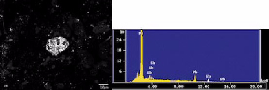
Secondary electron image of bullet fragment (left) and energy dispersive spectrometry (EDS) showing xray peaks for lead (Pb) and antimony (Sb).
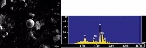
Secondary electron image of bullet fragment (left) and energy dispersive spectrometry (EDS) showing xray peaks for barium (Ba),lead (Pb) and antimony (Sb).
It should be noted that these samples were viewed and analysed manually for illustrative purposes only. In order for this to be accepted in a court of law as evidence there are strict guidelines that need to be followed in terms of exactly how and by whom samples are taken and a thorough chain of evidence has to be established from sampling through to analysis by an accepted laboratory. Typically this type of work is undertaken using automated, unattended software controls to eliminate human error and or bias.